Xrays are part of the naturally occurring electromagnetic spectrum. They are invisible to the human eye.
Xray art, and Xray flora fine art ‘photography’ is practiced by a handful of artists, the earliest being Dr. Dane Tasker, a radiologist who published his images of flowers in the early ’30’s.
Allan’s Xrays of floral images show in exquisite detail, the delicate unseen structures that can only be captured using xray equipment.
 |
 |
 |
|
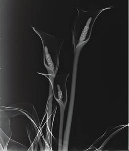
x-ray image before
|
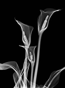
x-ray image after
|
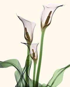
x-ray image after color process
|
THE TECHNIQUES:
White-On-Black Images
The White-on-Black images presented here are what we see with every-day medical imaging. The white areas represent the most dense, or thickest parts that the Xray beam passes through, resulting in less impact on the image sensor or film negative.
The resulting image is made up of white and dark areas corresponding to the varying densities of the subject being Xrayed. Indeed, there is an illusion of highlights as created in conventional studio lighting.
In floral Radiography the artist is challenged to demonstrate fine detail in both the delicate and less delicate structures of the object being Xrayed.
Black-On-White Images
The images presented in this form are simply inverted digitally similar to conventional photographic prints made from negatives.
Colour Images
The Xray images presented here start as either a black-on-white, or white-on-black image, then coloured digitally. This is a very exacting and time-consuming process. The result is an image that glows and radiates the beauty of its floral subject.
For more information about Allan Gill Photography, representing Dr. Gill, or how to obtain his art, please email: allangill38@gmail.com
No images may be duplicated without written permission from the artist.